- BioVector NTCC典型培養(yǎng)物保藏中心
- 聯(lián)系人:Dr.Xu, Biovector NTCC Inc.
電話:400-800-2947 工作QQ:1843439339 (微信同號)
郵件:Biovector@163.com
手機:18901268599
地址:北京
- 已注冊
Science:新熒光蛋白讓我們能觀察內(nèi)部器官
葉史瓦大學(xué)Albert Einstein醫(yī)學(xué)院研究人員首次研制出獨特的熒光蛋白,科學(xué)家借助該蛋白質(zhì)可清晰地觀察活體的內(nèi)部器官,因而擺脫外科手術(shù)的解剖刀和有輻射風(fēng)險的成像技術(shù)。
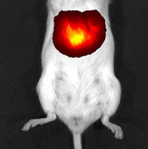
肝細(xì)胞表達(dá)iRFP蛋白
這一新探針在全身成像領(lǐng)域具有突破性意義,它能讓醫(yī)生在監(jiān)視腫瘤生長過程中不留下創(chuàng)傷,因而對癌癥治療效果的評估有積極作用。與人體掃描技術(shù)相比,熒光蛋白成像技術(shù)不涉及輻射風(fēng)險和對比劑。研究結(jié)果發(fā)表在7月17日 Nature Biotechnology期刊的網(wǎng)絡(luò)版上。
過去的20年,科學(xué)家嘗試了水母和珊瑚的各種有色熒光蛋白用于顯示細(xì)胞、細(xì)胞器和分子,但是熒光探針在哺乳動物體內(nèi)的使用卻存在挑戰(zhàn),動物血液中血紅蛋白能高效地吸收藍(lán)光、綠光、紅光以及其它波長的光。熒光蛋白的激發(fā)光和熒光蛋白“亮起來”后的發(fā)射光都會受到血紅蛋白的吸收干擾。
Einstein醫(yī)學(xué)院解剖與結(jié)構(gòu)生物學(xué)副教授 Vladislav Verkhusha博士為了解決這一問題,他的實驗室從細(xì)菌光敏色素(用于檢測光)提取出熒光蛋白,這一熒光蛋白(稱為iRFP)能吸收和發(fā)射電磁頻場的近紅外光,該頻段的光對于哺乳動物組織而言幾乎是透明的。
研究人員把熒光蛋白定位在肝臟中,這一器官由于血液含量高很難被觀察。
含有iRFP基因的腺病毒顆粒注入小鼠體內(nèi)。一旦載有iRFP基因的病毒感染肝細(xì)胞,感染細(xì)胞會表達(dá)該基因并合成iRFP蛋白質(zhì)。接著,小鼠暴露在近紅外光下,全身成像儀能觀察到發(fā)射熒光的肝臟。最早在感染后的第2天檢測出肝臟發(fā)熒光,5天后,熒光強度達(dá)到峰值。附加實驗表明,iRFP蛋白質(zhì)對哺乳動物沒有毒害作用。
論文的第1作者Grigory Filonov博士是 Einstein 醫(yī)學(xué)院 Verkhusha 實驗室的一個成員。他說,據(jù)研究所知,iRFP蛋白在活體肝臟顯示方面優(yōu)于其它熒光蛋白,它不僅產(chǎn)生非常明亮的圖像,而且長時期內(nèi)保持很高的穩(wěn)定性。我們相信它將在全身無創(chuàng)成像領(lǐng)域具有廣闊的應(yīng)用。
Filonov博士還說,與標(biāo)準(zhǔn)x-光和CT掃描相比,熒光蛋白成像沒有輻射風(fēng)險。此外,iRFP 蛋白成像很清晰,不需要對比劑,而核磁共振成像(MRI)需要吞下或注射對比劑以使得機體內(nèi)部結(jié)構(gòu)更清晰。(生物探索譯 Pobee)
生物探索推薦英文原文
Newly Developed Fluorescent Protein Makes Internal Organs Visible
Researchers at Albert Einstein College of Medicine of Yeshiva University have developed the first fluorescent protein that enables scientists to clearly "see" the internal organs of living animals without the need for a scalpel or imaging techniques that can have side effects or increase radiation exposure.
The new probe could prove to be a breakthrough in whole-body imaging -- allowing doctors, for example, to noninvasively monitor the growth of tumors in order to assess the effectiveness of anti-cancer therapies. In contrast to other body-scanning techniques, fluorescent-protein imaging does not involve radiation exposure or require the use of contrast agents. The findings are described in the July 17 online edition of Nature Biotechnology.
For the past 20 years, scientists have used a variety of colored fluorescent proteins, derived from jellyfish and corals, to visualize cells and their organelles and molecules. But using fluorescent probes to peer inside live mammals has posed a major challenge. The reason: hemoglobin in an animal's blood effectively absorbs the blue, green, red and other wavelengths used to stimulate standard fluorescent proteins along with any wavelengths emitted by the proteins when they do light up.
To overcome that roadblock, the laboratory of Vladislav Verkhusha, Ph.D., associate professor of anatomy and structural biology at Einstein and the study's senior author, engineered a fluorescent protein from a bacterial phytochrome (the pigment that a species of bacteria uses to detect light). This new phytochrome-based fluorescent protein, dubbed iRFP, both absorbs and emits light in the near-infrared portion of the electromagnetic spectrum- the spectral region in which mammalian tissues are nearly transparent.
The researchers targeted their fluorescent protein to the liver -- an organ particularly difficult to visualize because of its high blood content. Adenovirus particles containing the gene for iRFP were injected into mice. Once the viruses and their gene cargoes infected liver cells, the infected cells expressed the gene and produced iRFP protein. The mice were then exposed to near-infrared light and it was possible to visualize the resulting emitted fluorescent light using a whole-body imaging device. Fluorescence of the liver in the infected mice was first detected the second day after infection and reached a peak at day five. Additional experiments showed that the iRFP fluorescent protein was nontoxic.
"Our study found that iRFP was far superior to the other fluorescent proteins that reportedly help in visualizing the livers of live animals," said Grigory Filonov, Ph.D., a postdoctoral fellow in Dr. Verkhusha''''s laboratory at Einstein, and the first author of the Nature Biotechnology paper. "iRFP not only produced a far brighter image, with higher contrast than the other fluorescent proteins, but was also very stable over time. We believe it will significantly broaden the potential uses for noninvasive whole-body imaging."
Dr. Filonov noted that fluorescent-protein imaging involves no radiation risk, which can occur with standard x-rays and computed tomography (CT) scanning. And unlike magnetic resonance imaging (MRI), in which contrasting agents must sometimes be swallowed or injected to make internal body structures more visible, the contrast provided by iRFP is so vibrant that contrasting agents are not needed.
您正在向 biovector.net 發(fā)送關(guān)于產(chǎn)品 Science:iRFP,一種近紅外熒光蛋白,讓我們能觀察內(nèi)部器官 的詢問
- 公告/新聞


粒載體菌種細(xì)胞基因保藏中心http:www.biovector.net")
粒載體菌株細(xì)胞蛋白抗體基因保藏中心http:www.biovector.net")
